
FIGURE
Fig. 1
- ID
- ZDB-FIG-210323-34
- Publication
- Wang et al., 2021 - Py3-FITC: a new fluorescent probe for live cell imaging of collagen-rich tissues and ionocytes
- Other Figures
- All Figure Page
- Back to All Figure Page
Fig. 1
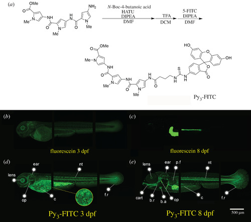
Py3-FITC can stain several tissues of zebrafish embryos. (a) Experimental procedure for the synthesis of Py3-FITC. Additionally, see electronic supplementary material, file S1 and figure S1. (b,c) Lateral view of fluorescein-stained zebrafish and (d,e) Py3-FITC-stained zebrafish embryos at (b,d) 3 dpf and (c,e) 8 dpf. Py3-FITC-stained lens, ear, opercle (op), notochord (nt) and cells distributed on the surface of the yolk sac and trunk region (c), fin rays (f.r.) 3 dpf. Jaw cartilage (cart), branchiostegal rays (b.r.), b.a. and pectoral fins (p.f.) were also stained 8 dpf. Scale bar in e, 500 µm. |
Expression Data
Expression Detail
Antibody Labeling
Phenotype Data
Phenotype Detail
Acknowledgments
This image is the copyrighted work of the attributed author or publisher, and
ZFIN has permission only to display this image to its users.
Additional permissions should be obtained from the applicable author or publisher of the image.
Full text @ Open Biol.