
Fig. 4
- ID
- ZDB-FIG-210216-12
- Publication
- Das et al., 2020 - Cortisol rapidly stimulates calcium waves in the developing trunk muscle of zebrafish
- Other Figures
- All Figure Page
- Back to All Figure Page
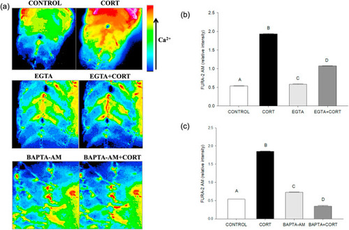
Fig. 4. Cortisol-mediated [(Ca2+)i] rise involves both intracellular and extracellular Ca2+. (a) Representative images of the developing trunk muscle (DTM) loaded with the ratiometric dye FURA-2AM and treated with either vehicle (CONTROL), cortisol (CORT), EGTA, BAPTA-AM or a combination of cortisol with EGTA (EGTA + CORT) or BAPTA-AM (BAPTA-AM + CORT). (b) A bar graph showing the ratiometric intensity at 30 s with extracellular calcium chelator EGTA either with or without cortisol. (c) A bar graph showing the ratiometric intensity at 30 s with an intracellular chelator BAPTA-AM either with or without cortisol. All bars represent mean ± SEM (n = 4–5 independent embryos); bars with different letters are significantly different; one way ANOVA; Holm Sidak post hoc test; p < 0.05. Abbreviations: Ethylene glycol-bis(β-aminoethyl ether)-N,N,N′,N′- tetraacetic acid (EGTA), 1,2-bis(o-aminophenoxy)ethane-N,N,N′,N′-tetraacetic acid-acetoxy methyl ester (BAPTA-AM). |
Reprinted from Molecular and Cellular Endocrinology, 520, Das, C., Faught, E., Vijayan, M.M., Cortisol rapidly stimulates calcium waves in the developing trunk muscle of zebrafish, 111067, Copyright (2020) with permission from Elsevier. Full text @ Mol. Cell. Endocrinol.