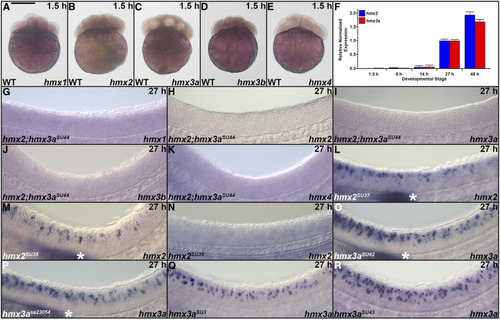
Expression of hmx genes in mutant zebrafish embryos and before the midblastula transition. (A–E, G–R) Lateral views of expression in whole embryos at 1.5 hpf (16 cells, A–E) or the spinal cord (G–R) at 27 hpf. (A–E) Animal pole, up. (G–R) Rostral, left; Dorsal, up. (L and M, O and P) White asterisk indicates expression in the lateral line primordium. None of the hmx genes are maternally expressed at 1.5 hpf, as assessed by in situ hybridization (A–E), and, in the case of hmx2 and hmx3a, quantitative RT-PCR on whole embryos (F). No maternal expression of hmx2 and hmx3a was detected and zygotic expression was not observed via quantitative RT-PCR until 14 hpf (F). hmx1 (G), hmx3b (J), and hmx4 (K) are not expressed in the spinal cord of hmx2;hmx3aSU44 deletion mutants. However, hmx1 and hmx4 were still expressed in the head, as shown in Figure 1 (data not shown), confirming that the in situ hybridization experiment had worked. We never detect expression of hmx3b in WT embryos at 27 hpf (see Figure 1). (H and I) As expected, given the deletion of the entire hmx3a coding sequence and all but the last 66 bp of hmx2 coding sequence in hmx2;hmx3aSU44 mutants (Figure 4), we did not detect any hmx2 (H) or hmx3a (I) transcripts in these mutants. (L and M) hmx2 mRNA does not exhibit nonsense-mediated decay (NMD) in hmx2SU37 or hmx2SU38 mutants. (N) In hmx2SU39 mutants, deletion of all but the first 84 and the last 60 bases of hmx2 coding sequence (Figure 4) generates a severely truncated hmx2 transcript that cannot be detected by our hmx2 ISH probe. Generation of a short ISH probe targeted to the predicted truncated transcript product of hmx2SU39 mutants also failed to detect hmx2 expression in these mutants (data not shown). (O–R) hmx3a mRNA does not exhibit NMD in hmx3aSU42 (O), hmx3asa23054 (P), hmx3aSU3 (Q), or hmx3aSU43 (R) mutant embryos. Bar, 280 µm (A–E), 50 µm (G–R).
|
