FIGURE

Figure 7

ID
ZDB-FIG-210113-7
Publication
England et al., 2020 - Hmx3a Has Essential Functions in Zebrafish Spinal Cord, Ear and Lateral Line Development
Other Figures
All Figure Page
Back to All Figure Page
Figure 7

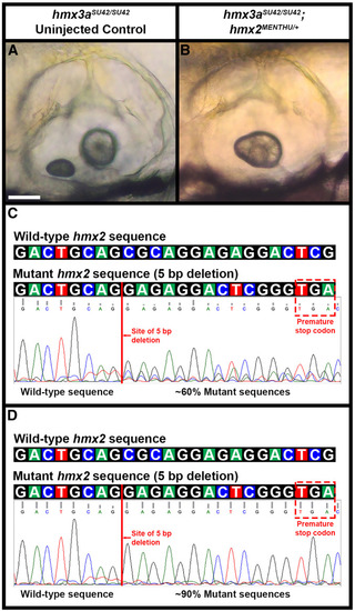
Phenotypic and genotypic analysis of embryos from an incross of hmx3aSU42/+ parents injected with hmx2MENTHU CRISPR reagents. (A and B) Lateral views of ear phenotypes at 4 day in live uninjected (A) and hmx2MENTHU CRISPR-injected (B) hmx3aSU42 homozygous mutant embryos. Rostral, left; Dorsal, top. The uninjected hmx3aSU42 homozygous mutant embryo has two normal otoliths in each ear (A). In contrast, the hmx2MENTHU CRISPR-injected hmx3aSU42 homozygous mutant embryo has fused otoliths in both ears. (C and D) Wild-type (top row) and hmx2MENTHU (bottom row) genomic sequences on top. Each colored box represents a specific nucleotide in the hmx2 coding sequence: A, green; C, blue; G, black; T, red. The hmx2MENTHU mutant sequence contains a 5 bp deletion (CGCAG, red line), which introduces a premature stop codon (red dashed box) 14–16 bases after the deletion. Sequencing traces (below) from individual embryos from an incross of hmx3aSU42/+ parents injected with hmx2MENTHU CRISPR reagents. In the injected embryos, hmx2MENTHU mutant sequences (with the 5 bp deletion) comprise ∼60% (C) to 90% (D) of all amplified sequences at this locus. Bar, 50 µm (A and B).

Expression Data

Expression Detail
Antibody Labeling
Phenotype Data
Fish:
Knockdown Reagent:
Observed In:
Stage: Day 4

Phenotype Detail
Acknowledgments
This image is the copyrighted work of the attributed author or publisher, and ZFIN has permission only to display this image to its users. Additional permissions should be obtained from the applicable author or publisher of the image. Full text @ Genetics