
FIGURE
Fig. 7-S1
- ID
- ZDB-FIG-200928-25
- Publication
- Bloch et al., 2020 - Non-thalamic origin of zebrafish sensory nuclei implies convergent evolution of visual pathways in amniotes and teleosts
- Other Figures
- All Figure Page
- Back to All Figure Page
Fig. 7-S1
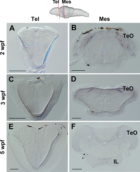
Expression of ert2Cre in the Tg(Dr830:ERT2CreERT2) juvenile brains.(A–F) Frontal sections of juvenile brains showing the telencephalic (Tel) and mesencephalic (Mes) areas. The levels of the frontal sections are indicated on the schematic drawing of a lateral view of the brain. In the 2 wpf (A and B), 3 wpf (C and D), and 5 wpf (E and F) juvenile brains, ert2Cre is expressed exclusively in the tectal area (B, D, and F). Abbreviations: IL, inferior lobe; Mes, mesencephalic area; Tel, telencephalic area; TeO, optic tectum. Scale bars = 100 µm (A–E); 200 µm (F). |
Expression Data
Expression Detail
Antibody Labeling
Phenotype Data
Phenotype Detail
Acknowledgments
This image is the copyrighted work of the attributed author or publisher, and
ZFIN has permission only to display this image to its users.
Additional permissions should be obtained from the applicable author or publisher of the image.
Full text @ Elife