FIGURE
            Fig. 3
- ID
 - ZDB-FIG-200928-17
 - Publication
 - Bloch et al., 2020 - Non-thalamic origin of zebrafish sensory nuclei implies convergent evolution of visual pathways in amniotes and teleosts
 - Other Figures
 - All Figure Page
 - Back to All Figure Page


                        Fig. 3


| 


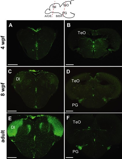
 Progression of GFP expression in the Tg(279A-GFP) brain during development.Frontal sections of 4 wpf (A and B), 8 wpf (C and D), and the adult (E and F) brains at the level of the telencephalon (A, C, and E) and PG (B, D, and F). Approximate antero-posterior levels are indicated in the schematic drawing on the top. At 4 wpf, there is no GFP+ fiber in the Dl (A) nor GFP+ cell around the PG (B). The GFP+ cells (presumably in the PGl) become obvious at 8 wpf (D), but their fiber labeling in the Dl is significantly weaker (C) in comparison to the adult (E). Abbreviations: Dl, lateral part of dorsal telencephalic area; PG, preglomerular complex; TeO, optic tectum. Scale bar = 200 µm.  | 


                        Expression Data


                        Expression Detail


                        Antibody Labeling


                        Phenotype Data


                        Phenotype Detail


                        Acknowledgments


This image is the copyrighted work of the attributed author or publisher, and
ZFIN has permission only to display this image to its users.
Additional permissions should be obtained from the applicable author or publisher of the image.
      Full text @ Elife