
Fig. 5-S1
- ID
- ZDB-FIG-200928-22
- Publication
- Bloch et al., 2020 - Non-thalamic origin of zebrafish sensory nuclei implies convergent evolution of visual pathways in amniotes and teleosts
- Other Figures
- All Figure Page
- Back to All Figure Page
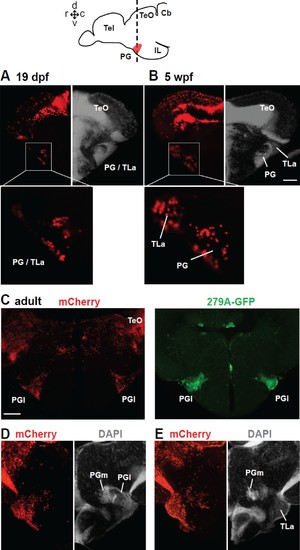
Distribution of mCherry+ cells in the PG following tamoxifen induction at 24 hpf in Tg(her5:ERT2CreERT2;βactin:lox-stop-lox-hmgb1-mCherry).A and B show frontal sections of juvenile brains, and C–E show of adult brains. The level of the frontal sections is indicated in the schematic drawing on the top. (A and B) mCherry+ cells at the level of PG at two different juvenile stages. The left half of the frontal section shows the distribution of mCherry (red), whereas the right half shows DAPI staining (grey) to illustrate the cytoarchitecture of the brain. At 19 dpf (A), the nucleus is labeled as ‘PG/TLa’, because they develop as a continuous structure and not easy to distinguish until around 5 wpf (B). (C) Adult brain sections at the level of PGl comparing the mCherry expression in Tg(her5:ERT2CreERT2;βactin:lox-stop-lox-hmgb1-mCherry) and GFP expression in Tg(279A-GFP). Abundant mCherry+ cells are found in the adult PGl, overlapping the location of GFP+ pallial projection neurons. (D and E) Adult brain sections showing more caudal levels than (C). The left half of the frontal section shows the distribution of mCherry (red), whereas the right half shows DAPI staining (grey) to illustrate the cytoarchitecture of the brain. E shows more caudal than D. The mCherry labelings are found throughout the PG, including the caudal PGl and PGm. Abbreviations: Cb, cerebellum; IL, inferior lobe; PG, preglomerular complex; PGl, lateral preglomerular nucleus; PGm, medial preglomerular nucleus; Tel, telencephalon, TeO, optic tectum; TLa, torus lateralis. Brain orientation: r, rostral; c, caudal; d, dorsal; v, ventral. Scale bars = 50 µm (B), 200 µm (C). |