
Fig 2
- ID
- ZDB-FIG-200829-103
- Publication
- Wilson et al., 2020 - A point mutation decouples the lipid transfer activities of microsomal triglyceride transfer protein
- Other Figures
- All Figure Page
- Back to All Figure Page
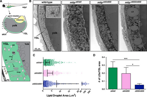
(A) (Top) Cartoon depicting the cross-sectional view of a 4 dpf zebrafish embryo. The YSL surrounds the yolk mass and serves as the embryonic digestive organ. The dashed box indicates the view expanded in the bottom panel and in panel B. (Bottom) Stored yolk lipids undergo lipolysis in yolk platelets (YP), presumably releasing free fatty acids into the YSL. These fatty acids are re-esterified in the ER bilayer to form TG, PL, and cholesterol esters. The lipids are packaged into B-lps in the ER with the help of Mtp and are likely further processed in the Golgi before being secreted into the perivitelline space (PS) and then circulation. (B) Representative transmission electron micrographs of the yolk and YSL from wild-type and |
| Fish: | |
|---|---|
| Observed In: | |
| Stage: | Day 4 |