IMAGE

Fig 2

ID
ZDB-IMAGE-200829-103
Source
Figures for Wilson et al., 2020
Image
Figure Caption

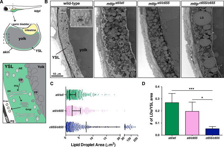
Fig 2 The opaque yolk phenotype results from the accumulation of aberrant cytoplasmic lipid droplets in the yolk syncytial layer.

(A) (Top) Cartoon depicting the cross-sectional view of a 4 dpf zebrafish embryo. The YSL surrounds the yolk mass and serves as the embryonic digestive organ. The dashed box indicates the view expanded in the bottom panel and in panel B. (Bottom) Stored yolk lipids undergo lipolysis in yolk platelets (YP), presumably releasing free fatty acids into the YSL. These fatty acids are re-esterified in the ER bilayer to form TG, PL, and cholesterol esters. The lipids are packaged into B-lps in the ER with the help of Mtp and are likely further processed in the Golgi before being secreted into the perivitelline space (PS) and then circulation. (B) Representative transmission electron micrographs of the yolk and YSL from wild-type and mttp mutants; dashed lines delineate the YSL region, mt = mitochondria, scale = 10 μm. (C) Quantification of lipid droplet size in mttp mutants, n ≥ 700 lipid droplets in 2 fish per genotype; mean +/- SD. (D) Quantification of the number of lipid droplets per YSL area, n = 7–9 YSL regions per genotype (3–5 regions per fish, 2 fish per genotype); mean +/- SD, Kruskall-Wallis with Dunn’s Multiple Comparison test, vs. mttpc655/c655, * p < 0.05, *** p < 0.001.

Figure Data
Acknowledgments
This image is the copyrighted work of the attributed author or publisher, and ZFIN has permission only to display this image to its users. Additional permissions should be obtained from the applicable author or publisher of the image. Full text @ PLoS Genet.