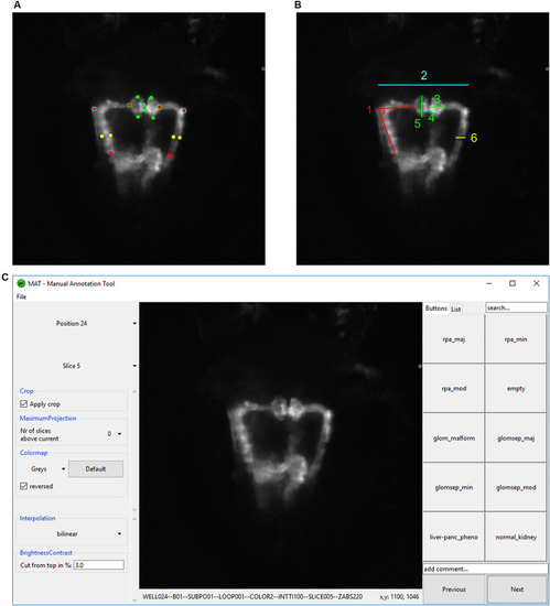
Analysis and annotation of pronephric phenotypes. (A,B) Quantitative measuring of morphological phenotypes of pronephroi. (A) Positions of 16 reference points are indicated that were manually assigned to each pronephros thumbnail image. (B) Illustration of calculated morphological parameters based on reference points in (A). Colors in (A,B) indicate which reference points in panel (A) were used for the calculation of morphological features in panel (B): (1) pronephric angle (only left side is shown – angleL), (2) tubular distance (tubDist), (3) glomerular height (only right side is shown – glomHeightR), (4) glomerular separation (glomSep), (5) glomerular width (only left side is shown – glomWidthL), and (6) tubular diameter (only right side is shown – tubDiamR). (C) Screenshot of software tool for manual annotation of large image datasets (https://doi.org/10.5281/zenodo.3367365). The manual annotation tool allows to browse, filter, auto-center, visualize and annotate complex multidimensional image datasets in an intuitive and blinded manner without any additional data pre-processing steps. In this study, the tool was used to assign up to 10 phenotypic categories to each acquired embryonic pronephros. Abbreviations: reduced pronephros angle major (rpa_maj), reduced pronephros angle minor (rpa_min), reduced pronephros angle moderate (rpa_mod), glomerular malformation (glom_malform), glomerular separation major (glomsep_maj), glomerular separation minor (glomsep_min), glomerular separation moderate (glomsep_mod), impaired liver pancreas area (liver-panc_pheno) and normal kidney (normal_kidney). Permission to reuse and Copyright: It is made available under a CC-BY-ND 4.0 International license.
|
