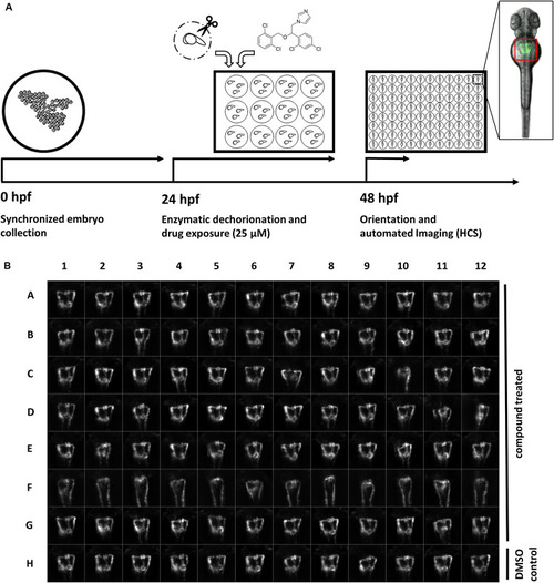
A screening workflow to score potential nephrotoxicity of approved drugs. (A) Schematic illustration of the drug toxicity screening pipeline. Synchronized embryos were obtained by collecting eggs from batch and pairwise crossings within a maximum interval of 30 min. At 24 h post fertilization (hpf) green fluorescent protein (GFP) positive embryos were enzymatically dechorionated using pronase and 20 embryos were transferred in each well of a 12-well plate. Different compounds were added to each well to a final concentration of 25 μM. At 48 hpf, and after 24 h drug exposure, embryos were washed, transferred into agarose filled microtiter plates, oriented within cavities generated with 3D-printed orientation tools and automatically imaged. The location of embryonic kidneys within image data was automatically determined using center of mass detection after thresholding of maximum z-projections. Center of mass coordinates were used to automatically crop maximum z-projections to thumbnails images with 257 × 257 pixels dimension [see also (B)]. Thumbnail images were used for all subsequent analysis steps. (B) Representative example of montage of pronephros thumbnail images, which was generated for each experimental 96-well plate. Each row was loaded with differently treated embryos. Row A-G show compound treated embryos, row F shows embryos with severely altered pronephros morphology, and row H shows embryos treated only with DMSO serving as plate internal negative controls. Permission to reuse and Copyright: It is made available under a CC-BY-ND 4.0 International license.
|
