
Fig. 4
- ID
- ZDB-FIG-200709-59
- Publication
- Kesavan et al., 2020 - Cell-fate plasticity, adhesion and cell sorting complementarily establish a sharp midbrain-hindbrain boundary
- Other Figures
- All Figure Page
- Back to All Figure Page
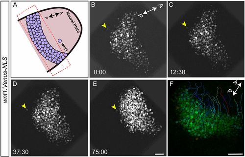
Cells sort at the MHB. To visualize cell sorting during MHB development in real time, embryos from the wnt1:Venus-NLS reporter line were mounted and imaged dorsally between 10.5 and 12 hpf, such that the neural plate and the neural keel stages were captured. Tissue sections spanning about 30 µm were chosen with a z-interval of 1 µm. Images were acquired at 2:30 (min:s) intervals. (A) Schematic representation marking the area of interest (box) and the orientation of the embryo used for time-lapse imaging. (B-E) wnt1:Venus-NLS- positive cells in the midbrain are seen undergoing morphogenetic process such as neural plate convergence and migration towards the anterior end. Importantly, one wnt1:Venus-positive cell (arrowheads) that was separated from the rest of the boundary cells showed active migration towards the group of other boundary cells. Time is in min:s. (F) Cell tracking showing the intermingling of cells with zig-zag movements and crossovers of tracks. Of the eight embryos analyzed by time-lapse imaging, active cell sorting was observed in five (62.5%). Scale bars: 40 µm. |