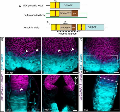
Initially overlapping expression domains segregate over time. (A) The knock-in strategy used for generating transgenic fish is schematized. A target site (Ts) located upstream of the open reading frame (ORF) of a gene of interest (GOI) is chosen. A bait plasmid is constructed by cloning the sequence upstream of the ORF, including the target site. The bait plasmid, sgRNA against the target site and Cas9 mRNA are injected into the embryo at the one-cell stage. The Cas9 protein creates double-stranded breaks at both Ts in the genomic locus and in the bait; this is followed by integration of the linearized bait plasmid. Only integration in the forward orientation will result in fluorescent reporter or CreERT2 expression. The primer pair (A+B) can be used to screen for and verify precise integration of the plasmid. The forward primer A is located outside the bait and the reverse primer B is located within the fluorescent reporter/CreERT2 sequence. (B) Live imaging was used to follow the expression of both mKate2 (membrane localized) and Venus, driven by the otx2b and gbx1 locus, respectively, at the various time points indicated. Overlapping expression boundaries were initially observed during the segmentation stages, i.e. between 10.5 and 16 hpf, and these segregated over time, based on changes in the presence of double-positive cells (mKate2 and Venus positive, indicated with arrowheads). The above-described phenotype was consistently observed in multiple embryos, and the total number of embryos (n) analyzed for each stage are: 10.5 hpf (n=7), 12 hpf (n=7), 16 hpf (n=4) and 30 hpf (n=4). (C) Snapshots from the time lapse imaging movie are shown; the overlapping otx2b-gbx1 region is indicated with a dotted rectangle. Time is shown in h:min. Scale bars: 20 µm. GOI, gene of interest; FP, fluorescent protein. The anterior-posterior axes of the embryos are marked with A-P and an arrow.
|
