FIGURE

FIG. 1.

ID
ZDB-FIG-200606-1
Publication
Keomanee-Dizon et al., 2020 - A versatile, multi-laser twin-microscope system for light-sheet imaging
Other Figures
All Figure Page
Back to All Figure Page
FIG. 1.

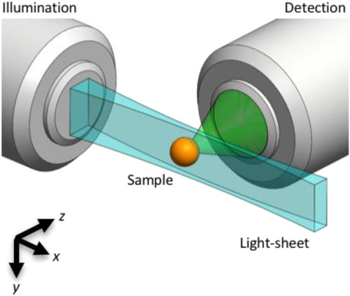
Light-sheet microscopy principle. A light-sheet (blue) can be created by dynamically scanning, along the y direction, a focused Gaussian beam that propagates in the x direction. The focusing is achieved via a low numerical aperture illumination lens. The fluorescence generated by the illuminated (x–y) plane is imaged (green) by an orthogonally positioned wide-field microscope. Axial (xy) sections of the 3D sample are captured either by scanning the sample (orange) through the stationary focal plane or by scanning the light sheet and detection focal plane through the stationary sample.

Expression Data

Expression Detail
Antibody Labeling
Phenotype Data

Phenotype Detail
Acknowledgments
This image is the copyrighted work of the attributed author or publisher, and ZFIN has permission only to display this image to its users. Additional permissions should be obtained from the applicable author or publisher of the image. Full text @ Rev. Sci. Instrum.