
Fig. 1
- ID
- ZDB-FIG-200521-11
- Publication
- Caetano-Lopes et al., 2020 - Unique and non-redundant function of csf1r paralogues in regulation and evolution of post-embryonic development of the zebrafish
- Other Figures
- All Figure Page
- Back to All Figure Page
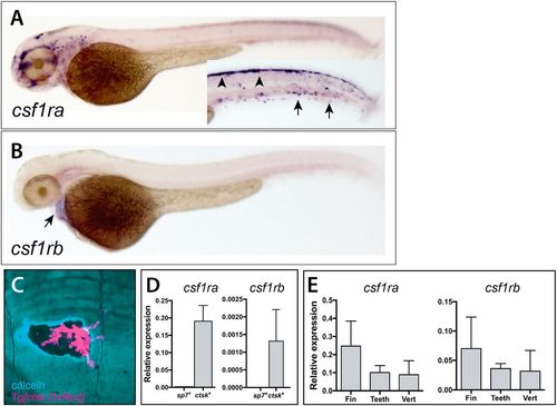
Divergent expression of csf1ra and csf1rb during zebrafish early development and in scales of the adult zebrafish. Whole-mount in situ hybridization of csf1ra (A) and csf1rb (B). (A) csf1ra is expressed in cells of the pigment and hematopoietic lineages at 2 dpf; inset shows the trunk in different focal plane with pigment cells precursors (arrowheads) and hematopoietic cells (arrows). (B) In contrast, csf1rb is expressed at only low levels in the heart (arrow) at 2 dpf. (C) Expression of Tg[cstk:DsRed] in osteoclasts localizes at areas of remodeling, as shown by increased calcein staining (blue) at the periphery of the remodeled pit. (D,E) Analysis of expression of csf1r paralogues in isolated cells and from adult skeletal tissues. (D) Expression of csfr1a and csf1rb in isolated adult osteoblasts (sp7+ cells) and osteoclasts (ctsk+ cells) from wild-type zebrafish. (E) Detection of both paralogues in isolated tissues [fin, teeth and vertebra (vert)]. |
| Genes: | |
|---|---|
| Fish: | |
| Anatomical Terms: | |
| Stage Range: | Long-pec to Adult |