
Fig. 3
- ID
- ZDB-FIG-200521-13
- Publication
- Caetano-Lopes et al., 2020 - Unique and non-redundant function of csf1r paralogues in regulation and evolution of post-embryonic development of the zebrafish
- Other Figures
- All Figure Page
- Back to All Figure Page
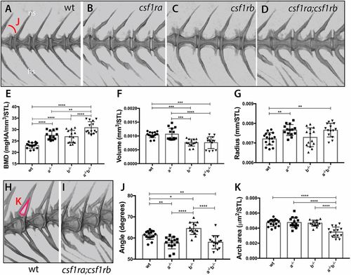
Loss of csf1r paralogue function leads to unique and shared phenotypes in the skeleton. MicroCT imaging and quantification of skeletal metrics of the spine of age-matched csf1r mutants and wild-type adult siblings. (A-D) MicroCT renderings of the lateral aspect of the spine showing overall morphology and integration of serial vertebrae. The neural spines (ns) and hemal spines (hs) are indicated. The measure used for the quantification of angle of the arch (J) is noted in A. (E-G,J,K) Quantification of vertebral shape and density in csf1r single and double mutants: (E) bone mineral density (BMD), (F) bone volume, (G) vertebral radius, (J) angle and (K) area of the neural arch. All measurements, with the exception of angle, were standardized to standard length (STL). (H,I) visualization of neural arch area in wild-type (H) and csf1ra; csf1rb mutant (I) zebrafish; measured area used for quantification of the arch area (K) is indicated in red in H. Data are mean±s.d. *P<0.05, **P<0.01, ***P<0.001, ****P<0.0001. |
| Fish: | |
|---|---|
| Observed In: | |
| Stage: | Adult |