FIGURE

FIGURE 5

ID
ZDB-FIG-200423-19
Publication
Carter et al., 2020 - Genetic Deletion of Zebrafish Rab28 Causes Defective Outer Segment Shedding, but Not Retinal Degeneration
Other Figures
All Figure Page
Back to All Figure Page
FIGURE 5

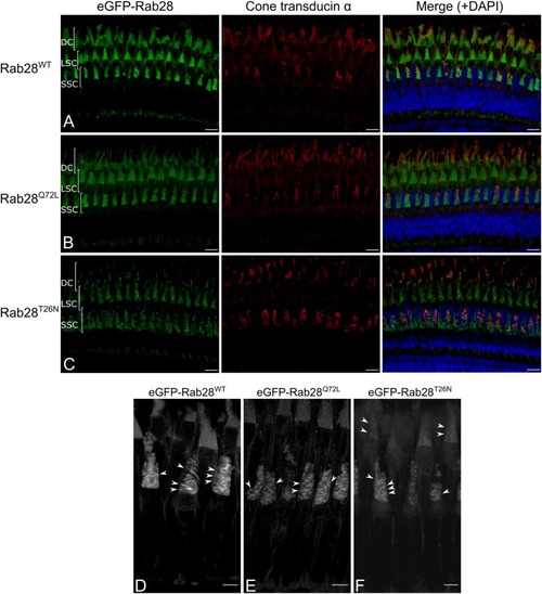
eGFP-Rab28 localization in 1 mpf zebrafish cone photoreceptors. (A–C) Representative confocal z-projections of eGFP-Rab28 localization in 1 mpf zebrafish cones. Cone OS are stained with anti-cone transducin alpha antibody. Each tier of photoreceptors is comprised of different classes of cone. DC: double cones; LSC: long single cones; SSC: short single cones. Scale bars 10 μm. For WT, Q72L and T26N eGFP-Rab28 reporters a total of 13, 11 and 11 retinas across at least six individuals were imaged, respectively. (D–F) Deconvolved, high resolution confocal z-projections of eGFP-Rab28 WT, Q72L and T26N mutant localization in 1 mpf cones. Arrowheads point to discrete bands present throughout the OS. Scale bars 5 μm.

Expression Data
Genes:
Antibody:
Fish:
Anatomical Term:
Stage: Days 30-44

Expression Detail
Antibody Labeling
Phenotype Data

Phenotype Detail
Acknowledgments
This image is the copyrighted work of the attributed author or publisher, and ZFIN has permission only to display this image to its users. Additional permissions should be obtained from the applicable author or publisher of the image. Full text @ Front Cell Dev Biol