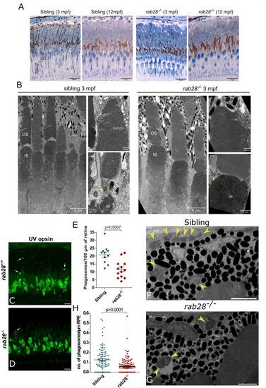
rab28 knockout zebrafish have reduced outer segment shedding, but normal retinal histology and ultrastructure. (A) Representative images of retinal histology in 3 and 12 mpf zebrafish, showing views of the photoreceptor layer in the central and peripheral retina in rab28ucd8 mutants and siblings. The overall structure and composition of the photoreceptor layer is grossly normal in both. Scale bars 20 μm (B) Representative transmission electron micrographs of 3 mpf zebrafish rab28ucd7 knockout and sibling retinas. Low magnification images show several cone photoreceptors, while high magnification images show examples of OS base and tips. Yellow arrows indicate ciliary basal body. OS: outer segment; IS: inner segment; m: mitochondria. Low magnification image scale bars 5 μm, high magnification scale bars 500 nm. (C,D) Confocal z-projections of rab28 mutant and sibling control retinas at 1 mpf, stained for UV opsin to label phagosomes (white arrows). Samples were collected 4 h after lights on. Scale bars 10 μm (E) Scatter plots of phagosome density in rab28 mutant and sibling retinas. Data are derived cryosections immunostained for UV and red opsins and cone transducin α. p-Value is derived from t-test. Error bars show SEM. Data are from 13 and 11 retinal z-projections from at least three individuals for mutants and siblings, respectively. (F,G) Representative TEM of RPE phagosomes in 15 dpf rab28 mutants and sibling controls. Yellow arrows indicate phagosomes. Samples were collected 4 h after lights on. Scale bars 2 μm. (H) Scatter plots of phagosome density in 15 dpf rab28 mutant and sibling retinas, derived from TEM. p-Value is derived from t-test. Error bars show SEM. Data are from three sibling and three mutant individuals.
|
