FIGURE

FIGURE 8

ID
ZDB-FIG-200423-22
Publication
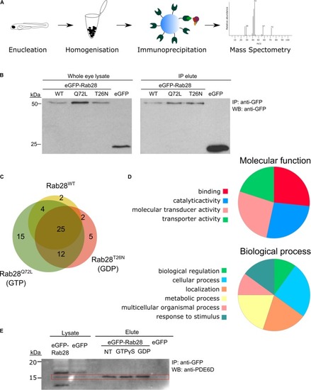
Carter et al., 2020 - Genetic Deletion of Zebrafish Rab28 Causes Defective Outer Segment Shedding, but Not Retinal Degeneration
Other Figures
All Figure Page
Back to All Figure Page
FIGURE 8

Rab28 interacts with multiple phototransduction proteins in the zebrafish eye. (A) Schematic of experimental workflow for Co-IP/MS of eGFP-Rab28 (B) Western blotting with an anti-GFP antibody, showing eGFP-Rab28 in whole eye lysate and elute after immunoprecipitation with anti-GFP beads. An untagged eGFP only control is also shown. (C) Venn diagram showing the number and overlap of significantly enriched (vs. GFP-only control) interacting proteins identified for each variant. (D) Pie charts showing gene ontology terms for proteins identified by mass-spectrometry following co-immunoprecipitation, which demonstrate a statistically significant interaction with any of the three variants. (E) Western blot of whole adult zebrafish eye lysate with an anti-PDE6D antibody, before and after IP of eGFP-Rab28. PDE6D band at 15 kDa mark (PDE6D mass: 17.4 kDa). Extra bands in the lysates are either post-translationally modified PDE6D, degradation products or non-specific binding by the antibody. IPs either received no treatment, GTPγS or GDP. eGFP only control is also shown.

Expression Data

Expression Detail
Antibody Labeling
Phenotype Data

Phenotype Detail
Acknowledgments
This image is the copyrighted work of the attributed author or publisher, and ZFIN has permission only to display this image to its users. Additional permissions should be obtained from the applicable author or publisher of the image. Full text @ Front Cell Dev Biol