FIGURE

Figure 5

ID
ZDB-FIG-200421-36
Publication
Adusumilli et al., 2020 - miR-7 Controls the Dopaminergic/Oligodendroglial Fate through Wnt/β-catenin Signaling Regulation
Other Figures
All Figure Page
Back to All Figure Page
Figure 5

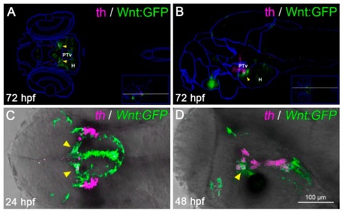
A subset of diencephalic Wnt-responsive cells express the dopaminergic (DA) marker tyrosine hydroxylase (TH). (A,B) Virtual anatomical reconstruction of a 72 hpf zebrafish brain, generated by ViBE-Z software, merging Wnt:GFP (green) and tyrosine hydroxylase (th, magenta) expression domains, compared with reference anatomical areas (blue lines). Virtual co-localizations (yellow arrowheads) are detected at the boundary between the ventral posterior tuberculum (PTv) and hypothalamus (H). (A,B) are dorsal and lateral views, respectively, with the anterior to the left, across the PTv/H boundary (section planes shown by white lines in the insets). (C,D) Co-localization of th-positive and Wnt-responsive areas (yellow arrowheads) is confirmed by dual color in situ hybridization in 24 and 48 hpf embryos. (C,D) are dorsal and lateral head views, respectively, with the anterior to the left.

Expression Data
Genes:
Fish:
Anatomical Terms:
Stage Range: Prim-5 to Protruding-mouth

Expression Detail
Antibody Labeling
Phenotype Data

Phenotype Detail
Acknowledgments
This image is the copyrighted work of the attributed author or publisher, and ZFIN has permission only to display this image to its users. Additional permissions should be obtained from the applicable author or publisher of the image. Full text @ Cells