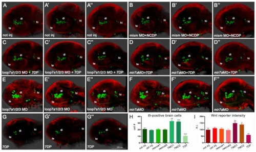
miR-7a negatively regulates Wnt signaling and diencephalic th-positive cells in the zebrafish brain (A–G’’): The knock-down of miR-7a activity by two independent morpholinos, targeting the immature and mature forms (loop7a1/2/3 MO, shown in E,E’,E’’, and mir7a MO, shown in F,F’,F’’) increases the amount of th-positive brain cells (in green) and the activity of a Wnt reporter (in red), compared with controls not injected (A,A’,A’’), co-injected with non-functional morpholino (mismMO) + non-functional miR-7a (NCDP) (B,B’,B’’), or co-injected (rescued) either with loop7a1/2/3 MO + 7DP (C,C’,C’’) or with miR-7a MO + 7DP (D,D’,D’’). On the contrary, over-expression of 7DP reduces the amount of th-positive brain cells and the activity of the Wnt reporter (G,G’,G’’). All pictures show the head region of 40 hpf embryos in lateral view, with the anterior to the left. te: telencephalon; di: diencephalon; hi: hindbrain. (H,I): Charts showing the th-positive cell counting (H), and the measure of Wnt reporter activity (I), expressed as fluorescence relative intensity (R.I.), in the seven conditions. Sample size n = 5 measures/condition; (*) p < 0.05; (**) p < 0.01; (***) p < 0.001.
|
