
Fig. S11
- ID
- ZDB-FIG-200306-133
- Publication
- Nimura et al., 2019 - Role of Reelin in cell positioning in the cerebellum and the cerebellum-like structure in zebrafish
- Other Figures
- All Figure Page
- Back to All Figure Page
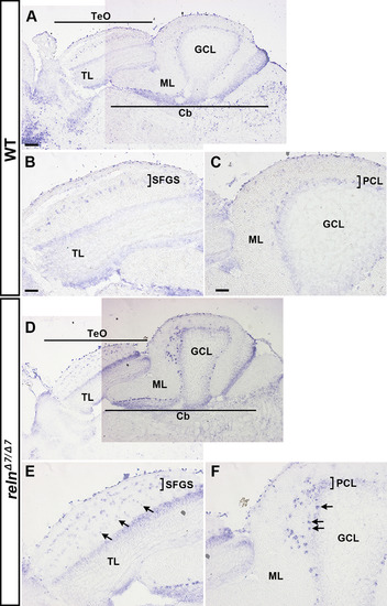
Ectopic type I neurons and Purkinje cells (PCs) in reln mutants. Sagittal sections from the brain of adult (159-dpf) WT (A-C, n= 2) and relnΔ7/Δ7 (D-F, n= 2) zebrafish were stained with an antisense riboprobe for Grid2 interacting protein a (grid2ipa). grid2ipa transcripts were detected in type I neurons located in the SFGS of the TeO, and in PCs in the PCL of the Cb in WT. Ectopic grid2ipa-expressing type I neurons and PCs were observed in the relnΔ7/Δ7 mutants (indicated by arrows). The abbreviations are described in the legend of Fig. 1. Scale bars: 100 μm in A (applies to A, D); 50 μm in B (applies to B, E); 50 μm in C (applies to C, F). |
Reprinted from Developmental Biology, 455(2), Nimura, T., Itoh, T., Hagio, H., Hayashi, T., Di Donato, V., Takeuchi, M., Itoh, T., Inoguchi, F., Sato, Y., Yamamoto, N., Katsuyama, Y., Del Bene, F., Shimizu, T., Hibi, M., Role of Reelin in cell positioning in the cerebellum and the cerebellum-like structure in zebrafish, 393-408, Copyright (2019) with permission from Elsevier. Full text @ Dev. Biol.