
Fig. 4
- ID
- ZDB-FIG-200306-119
- Publication
- Nimura et al., 2019 - Role of Reelin in cell positioning in the cerebellum and the cerebellum-like structure in zebrafish
- Other Figures
- All Figure Page
- Back to All Figure Page
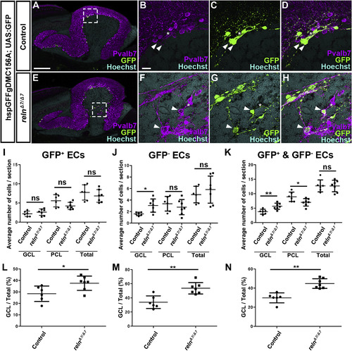
Ectopic eurydendroid cells (ECs) in reln mutants. (A–H) Ectopic ECs in relnΔ7/Δ7 mutants. Sagittal sections of adult (96-dpf) hspGFFgDMC156A; UAS:GFP zebrafish brains, which express GFP in ECs, harboring WT (control, n= 6) or homozygous reln mutant (relnΔ7/Δ7, n= 7) alleles were stained with anti-Pvalb7 (magenta), anti-GFP (green) antibodies, and Hoechst (nucleus, cyan). (B-D, F–H) High magnification images of the boxes in A and E. There are two types of ECs: GFP+(indicated by arrowheads) and GFP− (asterisks) ECs, both of which receive the Pvalb7+ axons of PCs (somata are surrounded by Pvalb7+ axons). Ectopic GFP+ and GFP− ECs were observed in the GCL of the reln mutant cerebellum (E–H). (I–K) GFP+ (I), GFP− (J), and total (J) ECs in the GCL, the PCL, or all layers (Total) of the controls and relnΔ7/Δ7 mutants were counted in every fourth section (18 total sections near the midline in each fish). Average numbers and standard deviations of ECs in the GCL, PCL, or all layers (Total) are shown in graphs. (L–N) Proportion of GFP+ (L), GFP− (M), or total (N) ECs in the GCL in controls and relnΔ7/Δ7 mutants. *p < 0.05; **p< 0.01; ns not significant (Welch’s t-test for GCL in J; Student’s t-test for I, PCL and Total in J, and K; Mann-Whitney test for L-N). Scale bars: 200 μm in A (applies to A and E); 40 μm in B (applies to B-D, F–H). |
| Genes: | |
|---|---|
| Antibody: | |
| Fish: | |
| Anatomical Terms: | |
| Stage: | Adult |
| Fish: | |
|---|---|
| Observed In: | |
| Stage: | Adult |
Reprinted from Developmental Biology, 455(2), Nimura, T., Itoh, T., Hagio, H., Hayashi, T., Di Donato, V., Takeuchi, M., Itoh, T., Inoguchi, F., Sato, Y., Yamamoto, N., Katsuyama, Y., Del Bene, F., Shimizu, T., Hibi, M., Role of Reelin in cell positioning in the cerebellum and the cerebellum-like structure in zebrafish, 393-408, Copyright (2019) with permission from Elsevier. Full text @ Dev. Biol.