
FIGURE
Fig. S7
- ID
- ZDB-FIG-200306-129
- Publication
- Nimura et al., 2019 - Role of Reelin in cell positioning in the cerebellum and the cerebellum-like structure in zebrafish
- Other Figures
- All Figure Page
- Back to All Figure Page
Fig. S7
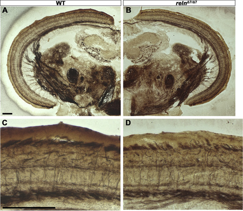
Layer structure of the optic tecum is not affected in reln mutants. Cross sections of the mesencephalon of adult WT (A, C, n= 2) and relnΔ7/Δ7mutant (B, D, n= 2) fish were stained by the Bielschowsky silver impregnation method, which visualizes neuronal fibers. (C, D) High magnification images of the tectum. Note that there was no difference in the neuronal fiber structure between the WT and the reln mutant tectum. Scale bars: 100 μm in A (applies to A, B) and C (applies to C, D). |
Expression Data
Expression Detail
Antibody Labeling
Phenotype Data
Phenotype Detail
Acknowledgments
This image is the copyrighted work of the attributed author or publisher, and
ZFIN has permission only to display this image to its users.
Additional permissions should be obtained from the applicable author or publisher of the image.
Reprinted from Developmental Biology, 455(2), Nimura, T., Itoh, T., Hagio, H., Hayashi, T., Di Donato, V., Takeuchi, M., Itoh, T., Inoguchi, F., Sato, Y., Yamamoto, N., Katsuyama, Y., Del Bene, F., Shimizu, T., Hibi, M., Role of Reelin in cell positioning in the cerebellum and the cerebellum-like structure in zebrafish, 393-408, Copyright (2019) with permission from Elsevier. Full text @ Dev. Biol.