
FIGURE
Fig. S2
- ID
- ZDB-FIG-200306-124
- Publication
- Nimura et al., 2019 - Role of Reelin in cell positioning in the cerebellum and the cerebellum-like structure in zebrafish
- Other Figures
- All Figure Page
- Back to All Figure Page
Fig. S2
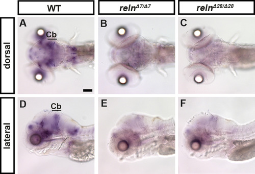
reln transcripts are severely decreased in reln mutants. Expression of reln in 5-dpf WT (A, D, n= 2), relnΔ7/Δ7 (B, E, n= 2), and relnΔ28/Δ28 (C, F, n= 2) larvae was analyzed by whole-mount in situ hybridization. Note that the reln expression was severely decreased in the reln mutants. Cb, cerebellum. Scale bars: 100 μm in A. |
Expression Data
Expression Detail
Antibody Labeling
Phenotype Data
Phenotype Detail
Acknowledgments
This image is the copyrighted work of the attributed author or publisher, and
ZFIN has permission only to display this image to its users.
Additional permissions should be obtained from the applicable author or publisher of the image.
Reprinted from Developmental Biology, 455(2), Nimura, T., Itoh, T., Hagio, H., Hayashi, T., Di Donato, V., Takeuchi, M., Itoh, T., Inoguchi, F., Sato, Y., Yamamoto, N., Katsuyama, Y., Del Bene, F., Shimizu, T., Hibi, M., Role of Reelin in cell positioning in the cerebellum and the cerebellum-like structure in zebrafish, 393-408, Copyright (2019) with permission from Elsevier. Full text @ Dev. Biol.