
Fig. S3
- ID
- ZDB-FIG-200306-125
- Publication
- Nimura et al., 2019 - Role of Reelin in cell positioning in the cerebellum and the cerebellum-like structure in zebrafish
- Other Figures
- All Figure Page
- Back to All Figure Page
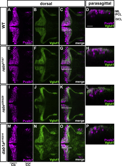
Cerebellum development is not affected in reln or dab1a mutant early-stage larvae. WT (A-D, n= 8), relnΔ7/Δ7 (E-H, n= 5), relnΔ28/Δ28 (I-L, n= 9),and dab1aΔ14/Δ14 (M-P, n= 11) larvae at 5 dpf were stained with anti-Pvalb7 (magenta) and anti-Vglut1 (green) antibodies. Dorsal views with anterior to the left (A-C, E-G, I–K, M-O). Sagittal section images at the level indicated by lines in C, G, K, and O are shown in D, H, L, and P. Typical images are shown. Note that no abnormalities in the development of Pvalb7+ PCs or Vglut1+ GC axons or in the layer formation were observed in the reln and dab1b mutants. CC, crest cerebellaris. The other abbreviations are described in the legend of Fig. 1. Scale bars: 100 μm in A (applies to A-C, E-G, I–K, M-O); 20 μm in D (applies to D, H, L, P). |
Reprinted from Developmental Biology, 455(2), Nimura, T., Itoh, T., Hagio, H., Hayashi, T., Di Donato, V., Takeuchi, M., Itoh, T., Inoguchi, F., Sato, Y., Yamamoto, N., Katsuyama, Y., Del Bene, F., Shimizu, T., Hibi, M., Role of Reelin in cell positioning in the cerebellum and the cerebellum-like structure in zebrafish, 393-408, Copyright (2019) with permission from Elsevier. Full text @ Dev. Biol.