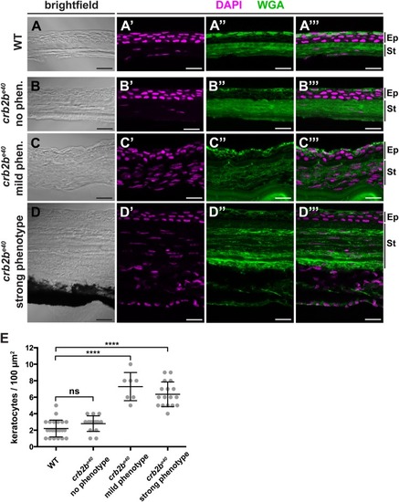
Loss of Crb2b-lf leads to an increase in the number of stromal keratocytes in old fish. (A–D‴). Brightfield (A,B,C,D), DAPI (magenta) and WGA (green) staining (A′–A″′,B′–B″′,C′-C″′,D′–D″′) of transverse cryosections of WT (A–A″′) and crb2be40 mutant (B–D″′) adult cornea. The stroma of crb2be40 mutants, which show an aberrant phenotype (C–D″′), appears to have more keratocyte nuclei (magenta) than mutants with no phenotype (B–B″′) or WT (A–A″′). Keratocyte nuclei were identified based on their flat and elongated morphology. Ep, corneal epidermis; St, corneal stroma. Scale bars: 5 µm. (E) Quantification of keratocyte nuclei in stromal tissue in WT versus crb2be40 mutant fish. The phenotypic categories correspond to the ones in A–D″′. Nuclei were counted from two separate 100 µm2 areas per adult fish in the central cornea, except for the ‘mild phenotype’ category due to lack of iris-free area at the centre of the cornea. Statistical significance was calculated by t-test (unpaired, with equal s.d., two-tailed). ****P<0.001; ns, not significant (P=0.0779).
|
