|
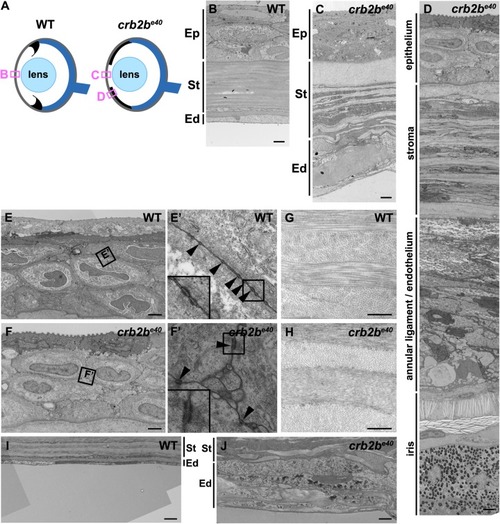
Loss of Crb2b-lf leads to corneal abnormalities in old fish. (A) Simplified schematic of a WT (left) and a crb2be40 (right) adult eye phenotype. Boxes denote regions of TEM images shown in B–D. (B–D) TEM transverse sections through the central cornea of adult WT (B) and crb2be40 fish (C,D) without (C) and with (D) iris expansion. (E–J) Higher magnification of the corneal layers in WT and mutant fish. Mutant corneal epidermal cells (F) do not appear different from their WT counterparts (E), and junctions (boxed regions in E,F) are visible in both (E′,F′, arrowheads). In the corneal stroma, collagen fibres are orthogonally layered both in the WT (G) and in the mutant (H). The endothelium is monolayered in WT (B,I), but appears as a multi-layered tissue in the mutant (C,J; labelled as annular ligament/endothelium in C), next to the elongated iris (D). Ep, corneal epidermis; St, corneal stroma; Ed, corneal endothelium. Scale bars: (B–F,I,J) 2 µm; (G,H) 0.5 µm.
|