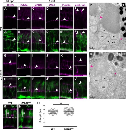
crb2be40 mutant PRCs have normal morphology and polarity. (A–L′) Immunostaining of transverse sections of WT (A–F′) and crb2be40 (G–L′) retinas in the Tg(bactin:mRas-EGFP) background at 51 hpf (A,A′,G,G′) and 5 dpf (B–F′,H–L′). Crb2a localises on the entire apical membrane in WT (A,A′) and crb2be40 mutants (G,G′) PRC precursors of this stage (arrowheads). At 5 dpf, Crb2a and aPKC localise to the IS (arrowheads) in WT (B,B′,C,C′) and in mutant cells (H,H′,I,I′). ZO-1 and F-actin localise to the outer limiting membrane (OLM; white arrowheads), both in WT (D–E′) and mutants (J–K′). A well-formed axoneme (white arrowheads) is detected in retinal sections in both WT (F,F′) and crb2be40 (L,L′) fish by acetylated tubulin antibody staining. White dashed lines indicate OLM. (M,N) Transverse retinal sections of WT (M) and crb2be40 (N) larvae at 3 dpf stained with Zpr-1 antibody to mark the cell body of double-cone PRCs (green) and phalloidin to mark the OLM (magenta). The solid white lines indicate IS height, the dashed white lines mark the position of the OLM. (O) Quantification of IS length (µm) in both WT and crb2be40 PRCs. IS length was measured from the level of the OLM to the base of the OS (see M,N). At least 20 cells from three independent retinas were measured. Statistical significance was calculated by t-test (unpaired, with equal s.d., two-tailed). ns, not significant (P=0.3053). (P,Q) Transmission electron micrographs of WT (P) and crb2be40 (Q) OSs at 3 dpf. The overall ultrastructure of cilia (magenta asterisks) and OS membrane discs (magenta arrowheads) is preserved in mutant PRCs. mt, mitochondrium. Scale bars: (A–N) 5 µm; (P,Q) 1 µm.
|
