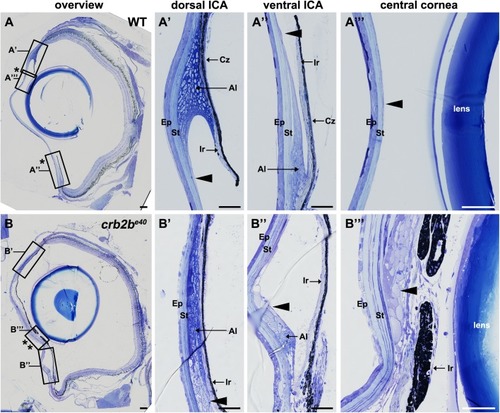
Loss of Crb2b-lf leads to an overgrowth of the iris in old fish. Toluidine Blue-stained transverse retinal sections of WT (A–A″′) and crb2be40 (B–B″′) adult zebrafish. (A–A″′) Overview of the whole WT eye (A) shows the normal appearance of the pupil (bordered by two asterisks). Higher magnification of WT dorsal (A′) and ventral (A″) iridocorneal angles shows the morphology of the iris, annular ligament cells and endothelial cells lining the ligament (arrowhead). A″′ shows the structure of the cornea, with the corneal endothelium cells being barely visible (A″′, arrowhead). (B–B″′) Overview of the whole eye of a mutant fish (B) demonstrates a decrease in pupil size (bordered by two asterisks) due to overgrowth of ICA tissue and an expansion of the iris towards the centre of the cornea (B″′). Multiple layers of cells line the posterior surface of the cornea (B″′, arrowhead). These cells appear to extend from the annular ligament (B′,B″ arrowheads). Ep, corneal epidermis; St, corneal stroma; Al, annular ligament; Ir, iris; Cz, ciliary zone. Scale bars: (A,B) 100 µm; (A′–A″′) and (B′–B″′) 50 µm.
|
