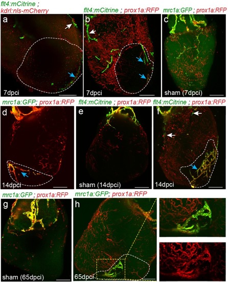
Figure 6 supplement 1.

Lymph-vasculogenesis represents the main mechanism of lymphatic formation after cardiac injury.

Injured area is outlined in all images. (a,b) seven dpci ventricles of Tg(kdrl:nls-mCherry);Tg(flt4BAC:mCitrine) (a) and Tg(prox1a:KalTA4-UAS:uncTagRFP); Tg(flt4BAC:mCitrine) (b) showing the opposite side of the hearts presented in Figure 6d,f, confirming the lack of connections between isolated LECs (blue arrows) and OFT originating ventricular lymphatics (white arrows). (c,e) Sham operated Tg(mrc1:EGFP);Tg(prox1a:KalTA4-UAS:uncTagRFP) hearts at seven dpci (c) (n = 3), and 65 dpci (g) (n = 2) and Tg(prox1a:KalTA4-UAS:uncTagRFP); Tg(flt4BAC:mCitrine) hearts at 14 dpci (e) (n = 3), showing no changes in lymphatic vasculature. (d,f) Opposite side of the hearts presented in Figure 6h,i, confirming the lack of connections between isolated LECs (blue arrows) and OFT originating ventricular lymphatics (white arrows). (h) Tg(mrc1:EGFP);Tg(prox1a:KalTA4-UAS:uncTagRFP) heart at 65 dpci showing increased lymphatic coverage in the injured vs. the uninjured areas of the ventricle (n = 4). Scale bars are 200 µm. All fish are 25–30 mm.

Expression Data

Expression Detail
Antibody Labeling
Phenotype Data

Phenotype Detail
Acknowledgments
This image is the copyrighted work of the attributed author or publisher, and ZFIN has permission only to display this image to its users. Additional permissions should be obtained from the applicable author or publisher of the image. Full text @ Elife