|
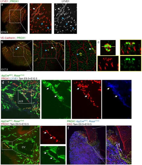
Morphological and lineage analysis of mouse cardiac lymphatics.(a,b) Whole mount images of embryonic hearts with isolated LEC clusters. (a) Co-staining with the nuclear lymphatic marker PROX1, and the membrane lymphatic marker, LYVE1, revealed that isolated LECs (blue arrow) do not connect to the main lymphatic vessel (white arrow, outline). (b) Isolated LEC clusters (blue arrows) were found as late at E17.5. Insets show increasing magnifications of a cluster that did not contact either VE-Cadherin+ blood ECs or developing lymphatic sprouts (white arrow). (c) An orthogonal view of the cluster highlighted in b) shows that blood vessels from deeper in the tissue do not connect. Scale bars are 200 µm. (d,e) Tissue sections through E11.5 ApjCreERT2,RosamTmG embryos dosed with tamoxifen at E9.5 and 10.5 showing labeling in the jugular lymph sacs (JLS) (green) (d), co-localized with PROX1 (red) and LYVE1 (blue) (inset, arrows). The cardinal vein (CV) is also labeled by mTmG (e) as well as a cluster of PROX1 labeled cell cluster budding from the CV (inset, arrows) (f) Inset is magnification of dashed box. Base of the aorta view of ApjCreER, RosamTmG embryos dosed with tamoxifen at E9.5 and 10.5 and analyzed at E15.5. Cre recombination is marked with GFP (blue), endothelial cells with VE-Cadherin (red) and lymphatics with PROX1 (green). Scale bars are 200 µm.
|