
Figure 1 supplement 2.
- ID
- ZDB-FIG-191230-1798
- Publication
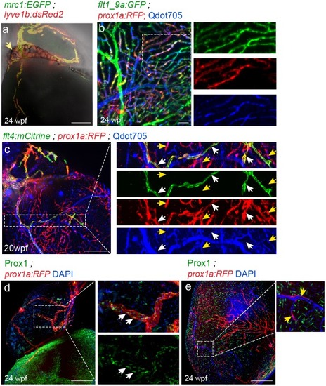
- Gancz et al., 2019 - Distinct origins and molecular mechanisms contribute to lymphatic formation during cardiac growth and regeneration
- Other Figures
- All Figure Page
- Back to All Figure Page
( |