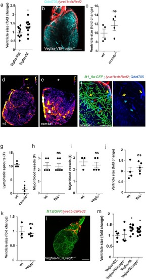
Ventricular size is increased following induction of Vegfaa-OE (nVegfaa-VEH = 9, nVegfaa-OE = 10, *p<0.01) (a), but remains unchanged in cxcr4a-/- mutants (c) (nwt = 4, ncxcr4a-/-=4). (b) Lymphatic sprouts (red) follow coronary vessels, labeled by intravascular injection of Qdot705 (cyan), in 12 wpf (fish size 19–22 mm) Vegfaa-OE hearts (n = 4) (d, e) Color-coded maps of vessel thickness show mis-patterned coronary arteries and absent major OFT vessels (e, asterisk) in cxcr4a -/-, as compared to wt siblings (d, yellow arrow). (f) cxcr4a -/- hearts display partially perfused blood coronary vessels (yellow arrows) following intravascular injection of Qdots705 at 25wpf (25–30 mm) (n = 4). (g) The number of ventricular lymphatic sprouts is reduced in 22 wpf (fish size 25–28 mm) Tg(flt1_9a_cFos:GFP);Tg(lyve1b:dsRed2) cxcr4a-/- hearts (nwt = 4, ncxcr4a-/-=4, *p<0.01). (h,i) Quantification of the number of major blood vessels in 19–23 wpf (fish size 25–30 mm) wt sibling and flt4 -/- hearts (h) (nwt = 5, nflt4 = 6) or 24 wpf (fish size 25–30 mm) wt sibling and vegfc+/- hearts (i) (nwt = 3, nvegfc +/-=5). (j,k) Quantification of ventricular size in 19–23 wpf (fish size 25–30 mm) wt sibling and flt4 -/- hearts (j) (nwt = 4, nflt4 = 5) and in 24 wpf (fish size 25–30 mm) wt sibling and vegfc +/- animals (k) (nwt = 3, nvegfc+/-=5). (l) 12 wpf (fish size 19–22 mm) Vegfaa-VEH;vegfc+/- fish in the background of Tg(fli1:EGFP);Tg(lyve1b:dsRed2). (m) Increased ventricular size is not reverted in 12 wpf (fish size 19–22 mm) Vegfaa-OE; vegfc+/- animals (nVegfaa-VEH = 5, nVegfaa-VEH;vegfc+/-=5, nVegfAa-OE-lymphatic vessel coverage=10 nVegfaa-OE;vegfc+/-lymphatic vessel coverage = 9, *p<0.01, relative to Vehicle treated sibling control). Error bars are 200 µm, mean ± S.E.M. All panels show anterior views.
Quantification of ventricular size in Vegfaa-OE, <italic>flt4 <sup>-/-</sup></italic>, <italic>vegfc<sup>+/ -</sup></italic> and <italic>cxcr4a <sup>-/-</sup></italic> hearts.
Expression Data
Expression Detail
Antibody Labeling
Phenotype Data
Phenotype Detail
Acknowledgments
This image is the copyrighted work of the attributed author or publisher, and
ZFIN has permission only to display this image to its users.
Additional permissions should be obtained from the applicable author or publisher of the image.
Full text @ Elife
Your Input Welcome
Thank you for submitting comments. Your input has been emailed to ZFIN curators who may contact you if
additional information is required.
Oops. Something went wrong. Please try again later.