
Fig. 6-S1
- ID
- ZDB-FIG-191021-6
- Publication
- Callahan et al., 2019 - Spinal V2b neurons reveal a role for ipsilateral inhibition in speed control
- Other Figures
- All Figure Page
- Back to All Figure Page
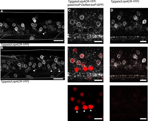
Anatomy of Tg(gata3:zipACR-YFP) expression.( A) and ( B) depict additional images of Tg(gata3:zipACR-YFP) expression in the full mediolateral extent of the spinal cord in one segment. White triangles mark putative CSF-cN appical extensions into the central canal. Noteably CSF-cN soma are not labeled with YFP. Scale bars = 20 μm. ( C) Spectrally deconvolved images of Tg(gata3:zipACR-YFP; gata3:loxP-DsRed-loxP:GFP), see Materials and methods. White (top) shows YFP emission and red (bottom) shows DsRed emission. Dorsal CSF-cN are noted with white triangles. CSF-cN somata are distinctly labeled with DsRed (BAC generated line) but not YFP (CRISPR generated line). Scale bar = 10 μm. ( D) Example spectral deconvolution of Tg(gata3:zipACR-YFP) showing negligible DsRed emission in control sample. Scale bar = 10 μm. |