
Fig. S1
- ID
- ZDB-FIG-190919-8
- Publication
- Vogrin et al., 2019 - Evolutionary Differences in the Vegf/Vegfr Code Reveal Organotypic Roles for the Endothelial Cell Receptor Kdr in Developmental Lymphangiogenesis
- Other Figures
- All Figure Page
- Back to All Figure Page
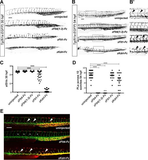
Analysis of primary and secondary sprouting in zebrafish embryos, Related to Figure 2 and STAR Methods. (A) Confocal micrographs of 30-hpf Tg(fli1a:EGFP) zebrafish embryos showing development of arterial intersegmental vessels (aISVs) in uninjected controls and embryos injected with mRNAs for soluble receptor constructs. Note the clearly abnormal vascular patterning in the embryos injected with mRNA for zFlt1-Fc and zKdrl-Fc. (B and B’) Confocal micrographs of 54-hpf Tg(fli1a:EGFP) zebrafish embryos showing development of parachordal LECs (PLs). Regions in the spotted rectangles are shown at higher magnification in B’ with arrowheads highlighting PLs, which are absent in embryos injected with mRNA for zFlt4(1-3)-Fc (asterisks). (C) Quantification of aISVs revealed a significant reduction following injection of mRNA for zFlt1-Fc (n=37 embryos) or zKdrl-Fc (n=28) compared to controls (n=46) (zFlt4-Fc n=47; zFlt4(1-3)-Fc n=27). **** denotes P<0.0001. (D) Quantification of PLs shows a reduction following injection of mRNA for zFlt4(1-3)-Fc, zFlt4-Fc or zKdrl-Fc compared to control (n=36, 29, 21 and 31, respectively; n=20 for zFlt1-Fc embryos). **** denotes P<0.0001. (E) Confocal micrographs of 54- hpf Tg(fli1a:nEGFP),Tg(-5.2lyve1b:dsRed) embryos showing PLs in uninjected controls (arrowheads), which are absent (asterisks) following injection of mRNA for zFlt4(1-3)-Fc, but present following injection of mRNA for zKdrl-Fc. Scale bars in A, B and E represent 100 m and apply to all images. |