
FIGURE
Fig. S3
- ID
- ZDB-FIG-190919-10
- Publication
- Vogrin et al., 2019 - Evolutionary Differences in the Vegf/Vegfr Code Reveal Organotypic Roles for the Endothelial Cell Receptor Kdr in Developmental Lymphangiogenesis
- Other Figures
- All Figure Page
- Back to All Figure Page
Fig. S3
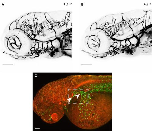
Microangiography shows no difference in cranial blood vessels in kdr mutants compared to wild-type siblings, Related to Figure 5. (A,B) Microangiography using 2000 kDa dextran fluorescein was used to detect blood vessel defects, however there were no obvious defects in blood vessel patterning at 5 dpf (n=4 mutant, n=6 wild-type sibling). (C) Confocal image of 36-hpf Tg(fli1a:nEGFP) transgenic embryo showing the region (boxed), containing the primary head sinus (arrow) and common cardinal vein (arrowhead), imaged in Figure 5I-L. Scale bars in A and B represent 100 m and in C 50 m. |
Expression Data
Expression Detail
Antibody Labeling
Phenotype Data
Phenotype Detail
Acknowledgments
This image is the copyrighted work of the attributed author or publisher, and
ZFIN has permission only to display this image to its users.
Additional permissions should be obtained from the applicable author or publisher of the image.
Full text @ Cell Rep.