
Fig. 6
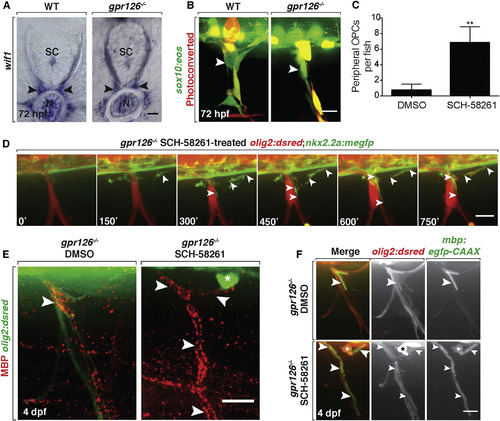
Peripheral OPCs Myelinate Spinal Motor Axons in gpr126Mutant Larvae (A) wif1 mRNA expression in WT and gpr126−/− larvae at 3 dpf shows MEP glia (arrowheads). SC, spinal cord; N, notochord. (B) Live images of photoconverted sox10:eos larvae at 3 dpf show Eos+MEP glia (arrowheads) in both WT and gpr126−/− larvae. (C) Mean ± SEM of peripheral OPCs in 3 dpf gpr126−/− larvae treated with DMSO or SCH-58261 from 36 hpf to 3 dpf. n = 8 DMSO, n = 6 SCH-58261. ∗∗p = 0.009. (D) Frames from a 15 h time-lapse movie of a olig2:dsred;nkx2.2a:megfp;gpr126−/− larva treated with SCH-58261 from 36 hpf to 3 dpf; 0′ is 57 hpf. Arrowheads mark a nkx2.2a+/olig2+ OPC ensheathing motor axons. (E) Images of MBP antibody (arrowheads) on peripheral nerves of 4 dpf olig2:dsred;gpr126−/− larvae treated with DMSO or SCH-58261 from 36 hpf to 3 dpf. Asterisk marks peripheral OPC. (F) Live images of 4 dpf WT and gpr126−/−;olig2:dsred;mbp:egfp larvae treated with DMSO or SCH-58261 from 36 hpf to 3 dpf. Asterisk marks olig2+ peripheral OPC with mbp+ membrane sheaths (arrowheads) around peripheral motor axons. Scale bars, 20 μm. |
| Genes: | |
|---|---|
| Antibody: | |
| Fish: | |
| Condition: | |
| Anatomical Terms: | |
| Stage Range: | Protruding-mouth to Day 4 |
| Fish: | |
|---|---|
| Condition: | |
| Observed In: | |
| Stage Range: | Protruding-mouth to Day 4 |