
Fig. S6
- ID
- ZDB-FIG-190814-8
- Publication
- Lin et al., 2019 - An Ectoderm-Derived Myeloid-like Cell Population Functions as Antigen Transporters for Langerhans Cells in Zebrafish Epidermis
- Other Figures
- All Figure Page
- Back to All Figure Page
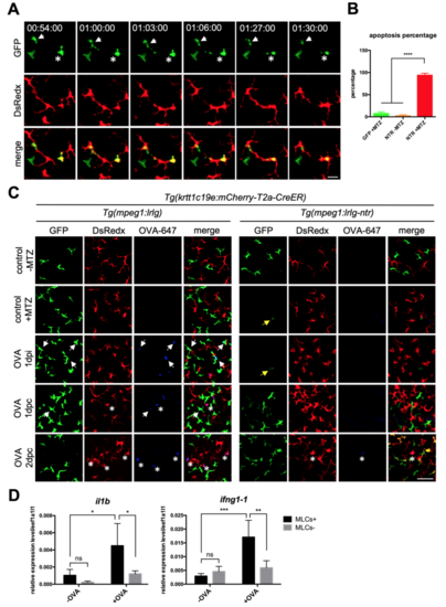
Depletion of MLCs impairs antigen uptake of cLCs. Related to Figure 6 (A) Live imaging on the scales of Tg(krtt1c19e:mCherry-T2aCreERT2;mpeg1:loxP-DsRedx-loxP-GFP-NTR) fish in L-15 culture media with 5 mM MTZ. White arrows and stars indicate two GFP-NTR+ MLCs undergo apoptosis and the dying cells are then phagocytized by DsRedx+ cLCs. Scale bar, 20μm. (B) Quantification of the percentages of apoptotic GFP+ MLCs in control Tg(krtt1c19e:mCherry-T2a-CreERT2;mpeg1:loxP-DsRedx-loxP-GFP) fish and experimental Tg(krtt1c19e:mCherry-T2a-CreERT2;mpeg1:loxP-DsRedx-loxPGFP-NTR) fish with or without 5 mM MTZ treatment (each group, n=4 fish). (C) Confocal images of the epidermis of control Tg(krtt1c19e:mCherry-T2aCreERT2;mpeg1:loxP-DsRedx-loxP-GFP) and experimental Tg(krtt1c19e:mCherry-T2a-CreERT2;mpeg1:loxP-DsRedx-loxP-GFP-NTR) fish (each group, n=4) after MTZ and OVA-647 treatments. Yellow arrows indicate few remaining MLCs in experimental fish after MTZ treatment. White arrows and stars show the colocalization of OVA-647 (blue) with MLCs (green) and cLCs (red) respectively. After depletion of MLCs (green), OVA-647 (blue) signals are significantly reduced in cLCs (red). Scale bar, 50μm. (D) Relative expression of il1b and ifng1-1 in whole fish epidermis at 2 dpc. In control fish (MLCs+, n=4), OVA-647 treatment induces significant increases of il1b and ifng1-1 expressions (black bar), whereas OVA-647-induced il1b and ifng1-1 expressions are dramatically reduced in the MLCs depleted fish (MLCs-, n=4) (grey bar). Data are represented as mean ± SD, *P<0.05, **P<0.01, ***P<0.001, ****P<0.0001. |
Reprinted from Developmental Cell, 49(4), Lin, X., Zhou, Q., Zhao, C., Lin, G., Xu, J., Wen, Z., An Ectoderm-Derived Myeloid-like Cell Population Functions as Antigen Transporters for Langerhans Cells in Zebrafish Epidermis, 605-617.e5, Copyright (2019) with permission from Elsevier. Full text @ Dev. Cell