
Fig. 5
- ID
- ZDB-FIG-190813-13
- Publication
- Lin et al., 2019 - An Ectoderm-Derived Myeloid-like Cell Population Functions as Antigen Transporters for Langerhans Cells in Zebrafish Epidermis
- Other Figures
- All Figure Page
- Back to All Figure Page
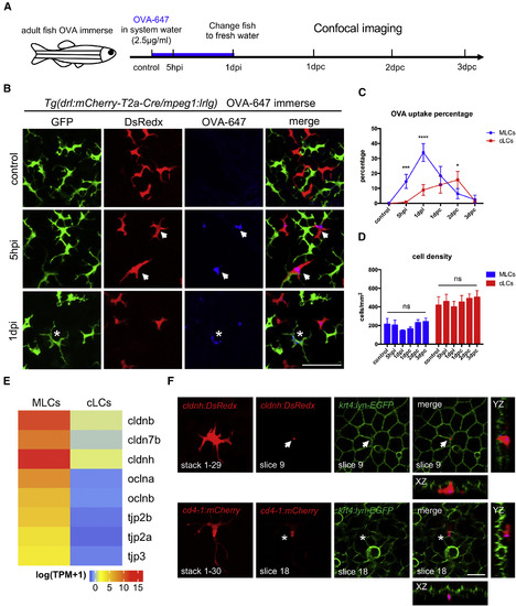
MLCs Sample Soluble Antigens from External Environment through Transepithelial Protrusions (A) A schematic diagram of experimental procedure of OVA-647 treatment of adult zebrafish. Whole fish are immersed in 2.5 μg/mL OVA-647 for 24 h (1 dpi) and then transferred to fresh system water without antigens for post-chase (pc) analysis. Images are captured at 5 h post immersion (hpi), 1 day post immersion (1 dpi), and 1 day post-chase (dpc), 2 dpc, and 3 dpc. (B) Confocal images of the epidermis of Tg(drl:mCherry-T2a-Cre;mpeg1:loxP-DsRedx-loxP-GFP) fish before (control), during (5 hpi), and after (1 dpi) OVA-647 immersion. Green, red, and blue signals represent cLCs, MLCs, and OVA-647, respectively. White arrows indicate OVA-647 in MLCs at 5 hpi, and white stars indicate OVA-647 in cLCs at 1 dpi. Scale bar, 50 μm. (C) Quantification of the percentages of OVA-647 uptake in MLCs (blue) and cLCs (red) at different time points (n = 4 fish). (D) Quantification of cell densities of MLCs (blue) and cLCs (red) at different time points (n = 4 fish). (E) Normalized average expression levels of tight junction genes [log(TPM+1)] in MLCs and cLCs from RNA-seq data (n = 4). (F) Confocal images of the epidermis in Tg(cldnh:DsRedx;krt4:lyn-GFP)and Tg(cd4-1:mCherry;krt4:lyn-GFP) fish after OVA-647 immersion. Upper panels show a typical transepithelial protrusion (white arrows) formed by MLCs (DsRedx+), which penetrates through the junction of keratinocytes and reaches the surface of krt4+ keratinocyte layer in XZ and YZ section views. Lower panels show normal protrusions (white star) formed by cLCs (mCherry+), which remain underneath krt4+keratinocyte layer. Scale bar, 10 μm. Data are represented as mean ± SD, ∗p < 0.05, ∗∗∗p < 0.001, ∗∗∗∗p < 0.0001. |
Reprinted from Developmental Cell, 49(4), Lin, X., Zhou, Q., Zhao, C., Lin, G., Xu, J., Wen, Z., An Ectoderm-Derived Myeloid-like Cell Population Functions as Antigen Transporters for Langerhans Cells in Zebrafish Epidermis, 605-617.e5, Copyright (2019) with permission from Elsevier. Full text @ Dev. Cell