
Fig. S5
- ID
- ZDB-FIG-190814-7
- Publication
- Lin et al., 2019 - An Ectoderm-Derived Myeloid-like Cell Population Functions as Antigen Transporters for Langerhans Cells in Zebrafish Epidermis
- Other Figures
- All Figure Page
- Back to All Figure Page
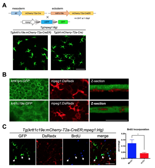
All mpeg1+ cells reside beneath the superficial krt4+ keratinocyte layer. Related to Figure 4, Figure 5 and Figure 6 (A) Generation double transgenic lines by crossing Tg(mpeg1:loxP-mIFP-loxPGFP) with Tg(krtt1c19e:mCherry-T2a-CreERT2) and Tg(drl:mCherry-T2a-Cre) fish. In these double transgenic lines, MLCs and cLCs are marked by GFP (lower panels). Scale bar, 50μm. (B) Confocal images of the epidermis of adult Tg(krt4:lyn-GFP;mpeg1:DsRedx) and Tg(krtt1c19e:GFP;mpeg1:DsRedx) fish. Z-section show that mpeg1+ cells (red) reside within krtt1c19e+ cells layer (green), which is beneath the krt4+ cells layer (green). Scale bar, 50μm. (C) Confocal images of Tg(krtt1c19e:mCherry-T2a-CreERT2;mpeg1:loxPDsRedx-loxP-GFP) fish, which were intraperitoneally injected BrdU at 1 day post OVA immersion (1dpi), then fixed at 2 day after injection (2dpc). White arrows indicate the incorporation of BrdU (blue) with MLCs (green). Quantification of BrdU incorporation percentages of MLCs and cLCs (n=3 fish). Scale bar, 50μm. Data are represented as mean ± SD, *P<0.05. |
Reprinted from Developmental Cell, 49(4), Lin, X., Zhou, Q., Zhao, C., Lin, G., Xu, J., Wen, Z., An Ectoderm-Derived Myeloid-like Cell Population Functions as Antigen Transporters for Langerhans Cells in Zebrafish Epidermis, 605-617.e5, Copyright (2019) with permission from Elsevier. Full text @ Dev. Cell