
Fig. 12
- ID
- ZDB-FIG-190618-47
- Publication
- Hanovice et al., 2019 - Regeneration of the zebrafish retinal pigment epithelium after widespread genetic ablation
- Other Figures
- All Figure Page
- Back to All Figure Page
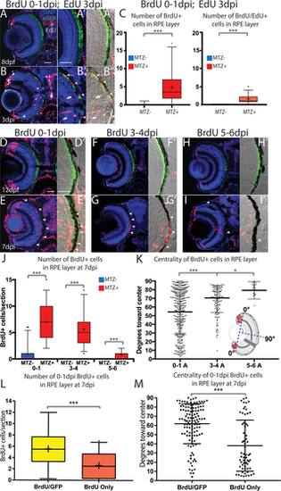
Proliferative RPE contributes to the regenerated RPE monolayer. (A-A”) Transverse sections from unablated larvae exposed to BrdU from 5-6dpf and pulsed with EdU for 2 hours before fixation at 8dpf. (B-B”) Transverse sections of ablated larvae exposed to BrdU from 0-1dpi and pulsed with EdU for 2 hours before fixation at 3dpi. (A’,B’) Magnified inset of BrdU/EdU. (A”,B”) Magnified inset of BrdU/EdU and DIC. Arrowheads in (B) highlight BrdU+ PRs that have integrated into the ONL. Arrow in (B’,B”) highlights a proliferative RPE cell, and arrowheads highlight unpigmented, previously-proliferative RPE-like cell in the injury site. (C) Quantification of BrdU/EdU+ and BrdU+ nuclei in the injury site. (D,E) Larvae exposed to BrdU 0-1dpi and fixed at 7dpi. (F-G) Larvae exposed to BrdU 3-4dpi and fixed at 7dpi. (H,I) Larvae exposed to BrdU 5-6dpi and fixed at 7dpi. (J) Quantification of the average number of BrdU+ cells per section. (K) Quantification of the location of individual BrdU+ cells relative to the center of the RPE. The line indicates the average location of BrdU+ cells, and the whiskers indicate standard deviation. (L,M) Quantification of BrdU+ cells that were labeled 0-1dpi within the RPE at 7dpi in ablated larvae. Analysis of eGFP+BrdU+and GFP-BrdU+ cells in the RPE reveal that most BrdU cells in the RPE are eGFP+ at 7dpi. (C) Quantification of the location of individual BrdU+ cells relative to the center of the RPE indicates that eGFP+BrdU+ cells tend to be located toward the center and eGFP-BrdU+ localize toward the periphery. Mann-Whitney U Test, * p<0.05, ** p<0.005, *** p<0.0005. Scale = 100μm. Dorsal is up and distal is left. Scale bar = 40um. |
| Fish: | |
|---|---|
| Condition: | |
| Observed In: | |
| Stage Range: | Day 6 to Days 7-13 |