
Fig. 5
- ID
- ZDB-FIG-190618-45
- Publication
- Hanovice et al., 2019 - Regeneration of the zebrafish retinal pigment epithelium after widespread genetic ablation
- Other Figures
- All Figure Page
- Back to All Figure Page
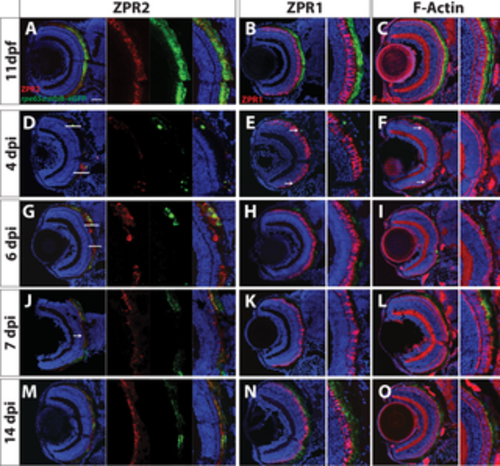
RPE regeneration initiates in the periphery and proceeds inward. Transverse sections of unablated larvae stained for the RPE marker ZPR2 (A), R/G cone photoreceptor marker ZPR1 (B) and F-Actin (C) at 11dpf. Ablated eyes stained for ZPR2 (D,G,J,M), ZPR1 (E,H,K,N), and Phalloidin (F,I,L,O) at 4, 6, 7 and 14dpi. Green = eGFP, blue = nuclei, red = marker. eGFP+ RPE appears in the periphery at 4dpi (marked by arrows in D-F). As regeneration proceeds, eGFP+ RPE extends further toward the eye center, and the leading tip of the regenerated monolayer often consists of both immature and mature RPE (ZPR2+/eGFP- cells in G). PR morphology appears to recover in the periphery proximal to regenerated RPE. By 7dpi, ZPR2+ RPE is present throughout the RPE (J), and PR morphology begins to recover in the central injury site (K,L). By 14dpi, mature eGFP+/ZPR2+ RPE cells are present throughout the RPE (M), and PR morphology further improves in the central retina (N,O). Dorsal is up and distal is left. Scale bar = 40μm. |
| Antibodies: | |
|---|---|
| Fish: | |
| Condition: | |
| Anatomical Terms: | |
| Stage Range: | Day 6 to Days 14-20 |
| Fish: | |
|---|---|
| Condition: | |
| Observed In: | |
| Stage Range: | Day 6 to Days 14-20 |