
Fig. 2
- ID
- ZDB-FIG-190618-44
- Publication
- Hanovice et al., 2019 - Regeneration of the zebrafish retinal pigment epithelium after widespread genetic ablation
- Other Figures
- All Figure Page
- Back to All Figure Page
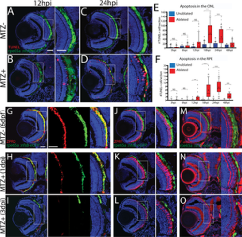
Ablation of the RPE leads to degeneration of underlying photoreceptors. (A-D) Transverse cryosections stained for TUNEL (red). Compared to untreated (A,C) larvae, ablated RPE were disrupted by 12hpi (B), and TUNEL+ cells appeared throughout the RPE and ONL at 24hpi (D). (E, F) Quantification of TUNEL+ cells/section in the RPE (E) and ONL (F) revealed a significant increase in the RPE by 12hpi and in the ONL by 18hpi. Significance determined using Mann-Whitney U test. * p≤0.05, ** p<0.005, *** p<0.0005. (G-I) Transverse sections of unablated 6dpf larvae stained for ZPR2 (G), ZPR1 (J), and F-Actin (M) (red). By 1dpi, ZPR2 is disrupted in a similar manner to eGFP (H), and ZPR1+ cones appear swollen and degenerated (K), and photoreceptor outer segment cytoskeletons become disorganized (N). By 3dpi, ZPR2 signal is absent from the central injury site (I) and PR morphology is notably degraded (L,O). Green = eGFP, blue = nuclei. Dorsal is up and distal is left. Scale bar = 40μm. |
| Antibodies: | |
|---|---|
| Fish: | |
| Condition: | |
| Anatomical Terms: | |
| Stage Range: | Day 6 to Days 7-13 |
| Fish: | |
|---|---|
| Condition: | |
| Observed In: | |
| Stage Range: | Day 6 to Days 7-13 |