
FIGURE
Fig. 8
- ID
- ZDB-FIG-190618-40
- Publication
- Hanovice et al., 2019 - Regeneration of the zebrafish retinal pigment epithelium after widespread genetic ablation
- Other Figures
- All Figure Page
- Back to All Figure Page
Fig. 8
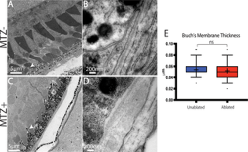
TEM analysis of regenerated RPE. (A,B) TEM images of unablated 19dpf and (C,D) 14dpi eyes (C) Organized photoreceptor outer segments are visible in the ablated photoreceptor layer, and a regenerated RPE is present. (E) Quantification of BM thickness. Student’s T-test reveals that BM thickness is not significantly different in ablated larvae * p<0.05. (MTZ- n = 3 eyes, 81 measurements; MTZ+ n = 3, 81 measurements). |
Expression Data
Expression Detail
Antibody Labeling
Phenotype Data
| Fish: | |
|---|---|
| Condition: | |
| Observed In: | |
| Stage Range: | Day 6 to Days 14-20 |
Phenotype Detail
Acknowledgments
This image is the copyrighted work of the attributed author or publisher, and
ZFIN has permission only to display this image to its users.
Additional permissions should be obtained from the applicable author or publisher of the image.
Full text @ PLoS Genet.