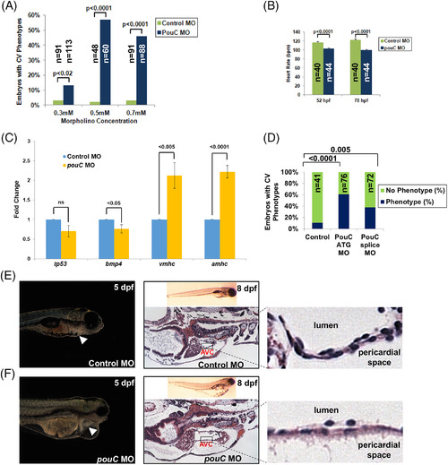
pouC knockdown impairs cardiac morphogenesis. A: Zebrafish embryos were injected with the indicated concentration of control or pouC MO and scored for a cardiovascular phenotype, which included pericardial edema, sluggish heart rate, abnormal looping, or impaired contraction. B: Heart rate was measured in control and pouC morphant zebrafish at 52 and 78 hpf. C: Zebrafish embryos were injected with control or pouC ATG MO. Total embryo RNA was isolated, and cDNA was prepared for qRT‐PCR analysis using the indicated primer sets. D: Zebrafish embryos were injected with control, pouC ATG, or pouC splicing MO. Cardiovascular phenotypes were assessed at 48 hpf. E: The left image shows a control zebrafish at 5 dpf with the heart indicated by a white arrowhead. The right image demonstrates a control zebrafish at 8 dpf in whole‐mount (top) and a section through the AVC with a zoomed view shown at right. F: Same as in (E) for a pouC morphant zebrafish. Note the decreased cellularity of the AVC region in pouC morphant embryos.
|
