
Fig. 3
- ID
- ZDB-FIG-190530-5
- Publication
- Bhakta et al., 2018 - pouC regulates expression of bmp4 during atrioventricular canal formation in zebrafish
- Other Figures
- All Figure Page
- Back to All Figure Page
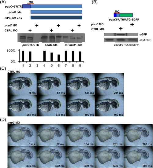
pouC knockdown perturbs cardiac function. A: in vitro transcription‐translation assay using reticulocyte lysate. Each of the indicated mRNA templates was incubated with no MO, control (CTRL) MO, or a pouC MO that binds to a region that overlaps the start codon. 35S‐labeled protein was resolved by sodium dodecyl sulfate‐polyacrylamide gel electrophoresis and exposed to film. Band quantitation (bottom) is depicted as a percentage intensity compared with untreated mRNA template. B: Zebrafish embryos were injected with the indicated pouC 5′UTR‐EGFP fusion mRNA alone or with CTRL MO or pouC MO. Embryo extracts were probed with the indicated antibodies by Western blotting. C: Video was captured at 78 hpf for a zebrafish embryo injected with control (CTRL) MO at the one‐cell stage. One cardiac cycle is shown with the indicated time stamp for each captured video frame. D: Same as in (C) for a 78 hpf zebrafish embryo injected with pouC MO. A slowed heart rate is indicated by the length of the cardiac cycle compared with the control embryo. Inefficient contraction, pooling of blood, and pericardial edema are also evident in the video frames. |
| Fish: | |
|---|---|
| Knockdown Reagent: | |
| Observed In: | |
| Stage: | Protruding-mouth |