
FIGURE
Fig. S3
Fig. S3
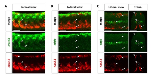
Characterization of the sclerotome. Wild-type embryos at 24 hpf were co-labeled with neural crest markers crestin (A, green), mitfa (B, green), or the muscle pioneer marker eng2 (C, green), with nkx3.1 (red). nkx3.1-expressing dorsal sclerotome (short arrows), ventral sclerotome (long arrows), and sclerotome derived notochord associated cells (arrowheads) do not express either crestin, mitfa, or eng2. The notochord (n) is indicated by brackets in lateral views and dotted lines in transverse views. n = 15 embryos per staining. Scale bars: 50 μm. |
Expression Data
Expression Detail
Antibody Labeling
Phenotype Data
Phenotype Detail
Acknowledgments
This image is the copyrighted work of the attributed author or publisher, and
ZFIN has permission only to display this image to its users.
Additional permissions should be obtained from the applicable author or publisher of the image.
Full text @ PLoS Genet.