
Fig. S1
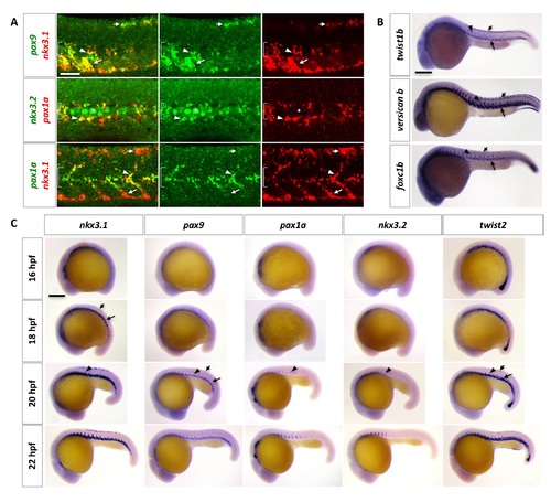
Characterization of the zebrafish sclerotome. (A) Double labeling of sclerotome markers in wild-type zebrafish at 24 hpf. nkx3.1 and pax9 expression overlaps in the dorsal sclerotome domain (short arrows), ventral sclerotome domain (long arrows), and sclerotome derived notochord associated cells (arrowheads). pax1a expression overlaps with nkx3.2 (middle panel) and nkx3.1 (bottom panel) only in the sclerotome derived notochord associated cells. The extent of the notochord is indicated by brackets. Non-specific labeling of notochord cells in nkx3.2 staining is indicated by asterisks. n = 60 embryos per staining. (B) Expression of twist1b, versican b, and foxc1b in wild-type zebrafish at 24 hpf. All 3 markers are expressed in the dorsal sclerotome (short arrows), the ventral sclerotome (long arrows), and sclerotome derived notochord associated cells (arrowheads). n = 15 embryos per staining. (C) Time course analysis of sclerotome marker expression in wild-type zebrafish between 16 hpf and 22 hpf. Expression of nkx3.1 and pax9 begins to appear in the ventral sclerotome domain (long arrows) at 16 hpf and 18 hpf, respectively. By 18 hpf, nkx3.1 is expressed in the dorsal sclerotome domain (short arrows). At 20 hpf, sclerotome derived cells (arrowheads) begin to “sprout” from the ventral domain, coinciding with the expression of pax1a and nkx3.2. Expression of nkx3.1 and pax9 is established in all three domains at this time. twist2 is also expressed in the sclerotome and has a similar timing and expression pattern as nkx3.1 and pax9. n = 30 embryos per staining. Scale bars: (A) 50 μm; (B, C) 200 μm. |