
Fig. 5
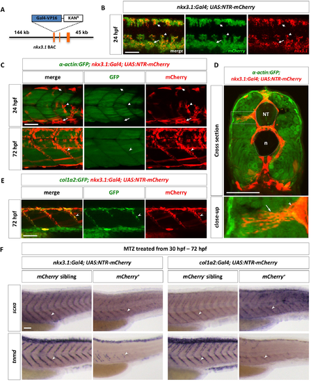
The sclerotome gives rise to axial tenocytes. (A) Schematic representation of the nkx3.1:Gal4 BAC reporter. A cassette containing Gal4-VP16 and Kanamycin resistance gene was recombined to replace the first coding exon of nkx3.1. (B) nkx3.1NTR-mCherry embryos were co-labeled with nkx3.1 (red) and ntr-mCherry (green) at 24 hpf. nkx3.1 and mCherry show overlapping expression in the dorsal sclerotome (short arrows), the ventral sclerotome (long arrows), and sclerotome derived notochord associated cells (arrowheads). n = 30 embryos. (C) The nkx3.1NTR-mCherry line (red) was crossed with the α-actin:GFP line (green) to label muscle cells. At 24 hpf, most mCherry+ cells, including dorsal sclerotome (short arrows), ventral sclerotome (long arrows), and sclerotome derived notochord associated cells (arrowheads), are not α-actin:GFP positive. Note that a few elongated mCherry+ muscle fibers are present in dorsal and ventral region of the somite. By 72 hpf, tenocytes (notched arrowheads) are found along the MTJ between adjacent somites. n = 5 embryos. (D) Cross section views of α-actin:GFP; nkx3.1NTR-mCherry embryos at 72 hpf show tenocytes (arrowheads) near at the medial edge of the somite extending long cellular projections (arrows) towards the surface of the body. The neural tube (NT) and notochord (n) are indicated by the dotted lines. An expanded view of an individual tenocyte in a boxed region is shown. n = 3 embryos. (E) Co-labeling of nkx3.1NTR-mCherry (red) with the col1a2:GFP (green) transgenic reporter at 72 hpf. mCherry+ tenocytes (notched arrowheads) along the MTJ also co-express col1a2:GFP. n = 15 embryos. (F) Embryos from nkx3.1NTR-mCherry or col1a2NTR-mCherry outcrosses were treated with metronidazole (MTZ) from 30 hpf to 72 hpf, and stained for the expression of scxa or tnmd. mCherry+ embryos showed markedly reduced numbers of tenocytes (notched arrowheads) as indicated by scxa or tnmd staining, compared to mCherry- sibling controls. n = 40 embryos per condition. Scale bars: 50 μm. |